Kelsey Foust
Senior Biology Major, Chemistry Minor
Laboratory/Field Proficiencies


Microscope Techniques
Microscope |
A skill that overlapped in almost every lab that I took was using a light microscope. A light microscope is an instrument that uses visible light and magnifying lenses in order to view small objects that are not visible to the naked eye or are extremely small. Light microscopes were used in my introductory Biology classes to veiw bacteria and small organisms such as Daphnia and were also used in my Histology class to view cross sections of mouse organs. While taking Histology, I learned that there were multiple types of microscopes that scientists use in order to view things that a light microscope may not be powerful enough to focus on. In Histology, I chose to research a Scanning Electron Microscope (SEM) to learn about what it is and some of its functions. Attached is a PowerPoint Presentation on the Scanning Electron Microscope.


Gel Electrophoresis
Electrophoresis|
During my time at WSSU, performing gel electrophoresis was one of my favorite skills that I acquired. Gel electrophoresis is a method used for the separation and analysis of macromolecules such as DNA and RNA based on their size and charge. During gel electrophoresis, an electrical current is sent through a porous medium ( polyacrylamide or agarose are commonly used) and DNA fragments are separated based on size and charge. Ultraviolet light is then used in order to analyze the results. Pictured at left is a gel electrophoresis run in my Biochemistry lab after PCR amplification. The goal of the experiment was to test for the D1S80 gene.


Spectrophotometer
Spectrophotometry |
Pictured is a spectrophotometer that was used in one of my Biochemistry lab experiments. Spectrophotometry is used in order to measure the reflection or transmittance properties of a material. In this lab, we monitored the activity of catalase on hydrogen peroxide based on the amount of light that was able to pass through the peroxide over a set amount of time.


In labs such as Organic Chemistry and Biochemistry, I learned about and demonstrated several chromatography techniques including affinity chromatography, and thin layer chromatography (TLC).In the affinity chromatography that I conducted in Biochemistry, the purpose of the experiment was to extract ConA from a jack bean meal mixture in order to determine the biological activity of Con A.In Organic Chemistry, we used thin layer chromatography for the majority of our experiments to either test the purity of a compound or if a reaction had run to completion.


In Microbiology, I learned about different types of bacteria, their life cycles, their effects on the body, and the different ways that they can be controlled. In lab, I learned a variety of techniques to distinguish between types of bacteria including characterizing bacteria by size and shape and using Gram staining to characterize bacteria based on its cell wall. During our final "Unknown lab", I combined the various identification techniques that I aquired over the semester to identify three strains of bacteria. I performed tests such as, Gram staining, Mannitol Salt Agar (MSA), Triple sugar ion (TSI), Citrate testing, Catalase testing, and Eosin Methylene Blue Agar (EMB) testing in order to identify my unknown bacterium.
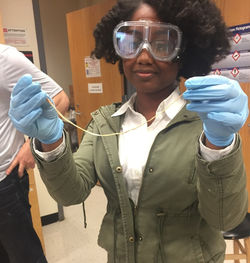 Nylon 6,6 |
|---|
Polymerization
In Organic Chemistry Lab, one of the most exciting experiments that we did was the preparation of Nylon 6,6 through polymerization. Nylon is a linear polyamide formed by a condensation reaction of bis-acyl chloride and a bis-amine. When we combined the two substances, aa stretchy polymer (nylon) was formed. We had a class competition to determine who could make the longer nylon polymer and my lab partner and I had the longest polymer, stretching almost the length of the Organic Chemistry lab! A video of our synthesized nylon is shown below.


